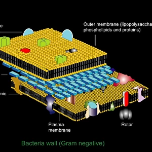
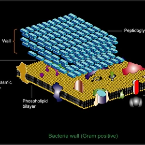
Bacterial cell wall comparison, artwork

![]()
Wall Art and Photo Gifts from Science Photo Library
Bacterial cell wall comparison, artwork
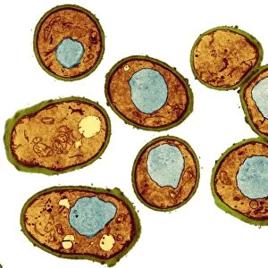
Bacterial cell wall comparison. Computer artwork comparing the structure of the cell wall from a gram-negative bacterium (left) with that of a gram-positive bacterium (right). Both forms have a cell membrane consisting of a phospholipid bilayer (blue), and then a layer of peptidoglycan (murein, orange) on the outside of this. The gram-negative cell wall has a thin peptidoglycan layer and then a second membrane (yellow), whereas the gram-positive one lacks the second membrane but has a much thicker peptidoglycan layer
Science Photo Library features Science and Medical images including photos and illustrations
Media ID 6322497
© PETER GARDINER/SCIENCE PHOTO LIBRARY
Bacteria Bacterial Bacteriology Bacterium Cell Wall Compared Comparing Comparison Gram Negative Gram Positive Label Labelled Labels Layer Membrane Membranes Micro Organism Micro Organisms Phospholipid Bilayer Phospholipids Micro Biology Microbiological
EDITORS COMMENTS
This artwork showcases a detailed comparison of bacterial cell walls, highlighting the distinct structures of gram-negative and gram-positive bacteria. The print presents a computer-generated image that beautifully illustrates the contrasting features between these two types of microorganisms. On the left side, we observe a gram-negative bacterium with its characteristic thin peptidoglycan layer followed by an additional outer membrane. This second membrane is depicted in vibrant yellow hues, emphasizing its presence as a unique trait in this type of bacteria. In contrast, on the right side, we see a gram-positive bacterium lacking this second membrane but compensating with an impressively thick peptidoglycan layer. Both forms share common elements such as a phospholipid bilayer (depicted in serene blue tones) forming their cell membranes and murein or orange-colored peptidoglycan layers surrounding them. These labels help us navigate through this intricate illustration and understand each component's role within these microscopic organisms. The white background enhances the clarity of this artwork while allowing viewers to focus solely on the structural differences being presented. With meticulous attention to detail and scientific accuracy, Science Photo Library has created an informative piece that merges artistry with biology seamlessly. This visually striking print serves as an invaluable resource for anyone interested in microbiology or bacteriology. It invites exploration into the fascinating world of bacterial anatomy while showcasing how diverse nature can be even at such minuscule scales.
MADE IN THE USA
Safe Shipping with 30 Day Money Back Guarantee
FREE PERSONALISATION*
We are proud to offer a range of customisation features including Personalised Captions, Color Filters and Picture Zoom Tools
SECURE PAYMENTS
We happily accept a wide range of payment options so you can pay for the things you need in the way that is most convenient for you
* Options may vary by product and licensing agreement. Zoomed Pictures can be adjusted in the Cart.