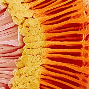
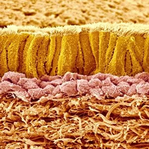
Eye anatomy, SEM

![]()
Wall Art and Photo Gifts from Science Photo Library
Eye anatomy, SEM
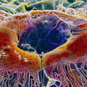
Eye anatomy. Coloured scanning electron micrograph (SEM) showing part of the ciliary body (blue) and iris (right) of an eye. The ciliary body is a ring-shaped structure that surrounds the iris and joins to ligaments that hold the lens in place behind the iris. It also contains the ciliary muscle that is contracted to alter the curvature of the lens and focus light on the retina at the back of the eye. Magnification: x20 when printed 10 centimetres wide
Science Photo Library features Science and Medical images including photos and illustrations
Media ID 6338269
© STEVE GSCHMEISSNER/SCIENCE PHOTO LIBRARY
Ciliary Body Colored False Colored Inside Internal Iris Ligament Ligaments Muscles Muscular Ocular Ophtalmological Ophthalmology Physiological Physiology Sight Tissue Vision False Coloured
EDITORS COMMENTS
This print from Science Photo Library offers a mesmerizing glimpse into the intricate world of eye anatomy. Through the lens of a scanning electron microscope (SEM), we are presented with a false-colored image that showcases the internal structures responsible for our vision. At first glance, our attention is drawn to the vibrant blue hues representing the ciliary body, which encircles and connects to ligaments holding the lens in place behind it. This ring-shaped structure plays a crucial role in adjusting the curvature of the lens through its contracted ciliary muscle, allowing us to focus light onto the retina at the back of our eyes. The level of detail captured by this SEM image is truly remarkable. Every muscular fiber and tissue within this delicate organ is brought to life, highlighting both its anatomical and physiological significance. It serves as a testament to how intricately designed our bodies are. As we delve deeper into understanding human biology, images like these become invaluable tools for researchers and medical professionals alike. They provide insights into ophthalmology and ocular health while fueling advancements in sight-related treatments. Whether you have an appreciation for scientific marvels or simply seek aesthetic beauty, this print invites you on an enlightening journey inside one of nature's most extraordinary creations –the human eye.
MADE IN THE USA
Safe Shipping with 30 Day Money Back Guarantee
FREE PERSONALISATION*
We are proud to offer a range of customisation features including Personalised Captions, Color Filters and Picture Zoom Tools
SECURE PAYMENTS
We happily accept a wide range of payment options so you can pay for the things you need in the way that is most convenient for you
* Options may vary by product and licensing agreement. Zoomed Pictures can be adjusted in the Cart.