False-col TEM of T4 bacteriophage in E. coli

![]()
Wall Art and Photo Gifts from Science Photo Library
False-col TEM of T4 bacteriophage in E. coli
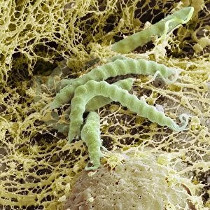
False colour transmission electron micrograph of bacteriophage T4 (virus infecting bacteria), 60 minutes after the injection of its viral DNA into the bacterium Escherichia coli. The phage, specific to E.coli, is secured to the surface by means of spidery tail fibres. The tail, a contrac- tile sheath, shortens to allow a syringe-like tube (visible below base plate of tail) to penetrate the cell membrane, emptying the DNA contents of the head into the bacterium. The capsid, a protein coat surrounding the DNA, remains outside the cell. Synthesis & assembly of the phage within the cell follows rapidly.Magnification:X32120 at 35mm size. Original is BW print m090/027
Science Photo Library features Science and Medical images including photos and illustrations
Media ID 6413052
© J. BROEK/BIOZENTRUM, UNIVERSITY OF BASEL SCIENCE PHOTO LIBRARY
Bacteriophage Bacteriophages Virology Viruses Micro Biology Virus
EDITORS COMMENTS
This print showcases a false-coloured transmission electron micrograph of the T4 bacteriophage infecting Escherichia coli, commonly known as E. coli. Taken 60 minutes after the injection of its viral DNA into the bacterium, this image provides a mesmerizing glimpse into the intricate world of virology. The T4 bacteriophage, specifically tailored to target E. coli, is firmly anchored to the bacterial surface by delicate spidery tail fibres. The tail possesses a remarkable ability to contract and shorten, enabling a syringe-like tube (visible below the base plate) to penetrate the cell membrane with precision. Through this process, the viral DNA contained within the phage's head is emptied into its host cell while leaving behind its protein coat called capsid. Magnified at an astonishing X32120 and captured on a black-and-white print m090/027 film, this photograph allows us to appreciate both the complexity and beauty inherent in microbiology research. It serves as a testament to our ever-expanding knowledge of viruses and their interactions with bacteria. With synthesis and assembly processes rapidly unfolding within E. coli after infection, this image offers just a snapshot of an ongoing battle between virus and host at microscopic scales. As we delve deeper into understanding these fascinating organisms through scientific exploration like this one from Science Photo Library's collection, we gain valuable insights that can aid in combating infectious diseases plaguing humanity today.
MADE IN THE USA
Safe Shipping with 30 Day Money Back Guarantee
FREE PERSONALISATION*
We are proud to offer a range of customisation features including Personalised Captions, Color Filters and Picture Zoom Tools
SECURE PAYMENTS
We happily accept a wide range of payment options so you can pay for the things you need in the way that is most convenient for you
* Options may vary by product and licensing agreement. Zoomed Pictures can be adjusted in the Cart.