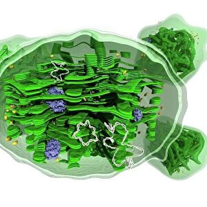
Liverwort leaf tissue, light micrograph

![]()
Wall Art and Photo Gifts from Science Photo Library
Liverwort leaf tissue, light micrograph
Liverwort leaf tissue. Light micrograph of the bifurcated tip of a leaf from a leafy liverwort (Lophocolea cuspidata). These leaves are extremely thin, consisting of one layer of cells full of chloroplasts. The cells are mainly hexagonal or pentagonal, and each cell has a peripheral layer of cytoplasm containing round chloroplasts (coloured dots) around a large central vacuole. Magnification: x195 when printed at 10 centimetres across
Science Photo Library features Science and Medical images including photos and illustrations
Media ID 6334267
© DR KEITH WHEELER/SCIENCE PHOTO LIBRARY
Cellular Chloroplast Chloroplasts Stem Thin Tissue Vacuole Vacuoles Cells Light Micrograph Light Microscope
EDITORS COMMENTS
This print showcases the intricate beauty of liverwort leaf tissue under a light microscope. The image captures the bifurcated tip of a leaf from a leafy liverwort species called Lophocolea cuspidata. These leaves are incredibly delicate, consisting of just one layer of cells that are brimming with chloroplasts. Under magnification, we can observe that the cells forming these leaves are predominantly hexagonal or pentagonal in shape. Each cell contains a peripheral layer of cytoplasm housing numerous round chloroplasts, which appear as vibrant colored dots throughout the image. Surrounding these cellular structures is a large central vacuole, adding to the overall visual complexity. Printed at 10 centimeters across and magnified 195 times, this photograph allows us to appreciate the remarkable details present within this botanical wonder. It provides an insight into the biological intricacies involved in plant growth and development. This stunning micrograph not only highlights nature's incredible diversity but also serves as a testament to the power of scientific imaging techniques. By capturing such breathtaking images, Science Photo Library continues its mission to inspire curiosity and foster appreciation for our natural world through their extensive collection of scientifically accurate visuals.
MADE IN THE USA
Safe Shipping with 30 Day Money Back Guarantee
FREE PERSONALISATION*
We are proud to offer a range of customisation features including Personalised Captions, Color Filters and Picture Zoom Tools
SECURE PAYMENTS
We happily accept a wide range of payment options so you can pay for the things you need in the way that is most convenient for you
* Options may vary by product and licensing agreement. Zoomed Pictures can be adjusted in the Cart.