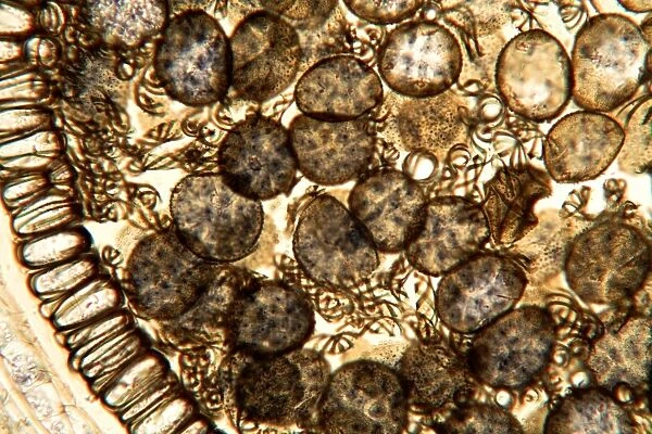
Liverwort spores, light micrograph
![]()

Wall Art and Photo Gifts from Science Photo Library
Liverwort spores, light micrograph
Liverwort spores, light micrograph. Transverse section through the sporangium of a liverwort (Pellia epiphylla). Part of the sporangiums outer wall is at left. The spherical spores take up most of the image. Between the spores are elongated cells with spiral thickening (elaters). When the sporangium splits open these elaters twist around as they dry and flick out the spores, which are then carried away by air currents. Magnification: x100 when printed at 10 centimetres wide
Science Photo Library features Science and Medical images including photos and illustrations
Media ID 6307793
© DR KEITH WHEELER/SCIENCE PHOTO LIBRARY
Bryophyte Bryophytes Bryophytic Cellular Cross Section Elater Elaters Internal Structure Liverwort Outer Wall Part Parts Plant Anatomy Re Production Reproductive Reproductive Part Reproductive Parts Sporangium Spore Spores Structural Tissue Transverse Cells Light Micrograph Light Microscope Section Sectioned
EDITORS COMMENTS
This print showcases the intricate world of liverwort spores, as seen through a light micrograph. The image reveals a transverse section of a liverwort sporangium belonging to the Pellia epiphylla species. On the left side, we can observe part of the sporangium's outer wall, while most of the frame is occupied by spherical spores. Interestingly, nestled between these spores are elongated cells with spiral thickening known as elaters. These specialized structures play a crucial role in dispersing the spores. As the sporangium splits open, these elaters dry out and twist around, creating tension that eventually propels them outward along with the spores. Once released into their surroundings, air currents carry them away to new locations where they can germinate and grow. The magnification used for this print is x100 when printed at 10 centimeters wide, allowing us to appreciate even finer details within this botanical marvel. This image provides valuable insights into both plant anatomy and reproductive processes in bryophytes like liverworts. Captured by Science Photo Library, this stunning photograph not only highlights nature's beauty but also serves as an invaluable resource for researchers and enthusiasts interested in cell biology, botany, and plant reproduction.
MADE IN THE USA
Safe Shipping with 30 Day Money Back Guarantee
FREE PERSONALISATION*
We are proud to offer a range of customisation features including Personalised Captions, Color Filters and Picture Zoom Tools
SECURE PAYMENTS
We happily accept a wide range of payment options so you can pay for the things you need in the way that is most convenient for you
* Options may vary by product and licensing agreement. Zoomed Pictures can be adjusted in the Cart.


