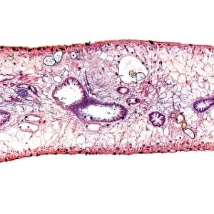
Moss reproductive parts, light micrograph

![]()
Wall Art and Photo Gifts from Science Photo Library
Moss reproductive parts, light micrograph
Moss reproductive parts, light micrograph. Longitudinal section through the antheridial cup (male reproductive parts) of a cord moss (Polytrichum commure). Inside the outer ring of leaves are the oval-shaped antheridia (dark pink). It is these structures that produce the antherozoids. Between the antheridia are multi-cellular hairs (paraphyses), which fill in the gaps and retain water for the antherozoids to swim in, to reach and fertilise a female moss plant and its eggs. Magnification: x45 when printed at 10 centimetres high
Science Photo Library features Science and Medical images including photos and illustrations
Media ID 6308409
© DR KEITH WHEELER/SCIENCE PHOTO LIBRARY
Antheridia Antheridium Bryophyte Bryophytes Bryophytic Cellular Internal Structure Longitudinal Moss Mosses Paraphyses Part Parts Plant Anatomy Re Production Reproductive Reproductive Part Reproductive Parts Structural Tissue Cells Light Micrograph Light Microscope Section Sectioned
EDITORS COMMENTS
This print showcases the intricate and fascinating world of moss reproductive parts. In this light micrograph, we are presented with a longitudinal section through the antheridial cup, which represents the male reproductive parts of a cord moss known as Polytrichum commure. The outer ring of leaves encloses oval-shaped structures called antheridia, depicted in a striking dark pink hue. These antheridia play a crucial role in producing antherozoids, tiny swimming entities responsible for fertilizing female moss plants and their eggs. To facilitate this process, multi-cellular hairs known as paraphyses fill in the gaps between the antheridia while retaining water for the antherozoids to swim in. With a magnification of x45 when printed at 10 centimeters high, this image provides us with a glimpse into the internal structure and anatomical details of these bryophytes. The white background accentuates every minute cellular detail captured by the light microscope used to create this stunning light micrograph. As we delve into plant biology and botany through this photograph from Science Photo Library, we gain insight into how nature's smallest organisms contribute to reproduction and ensure their survival. This visual representation serves as both educational material for those interested in plant anatomy and appreciation for the beauty found within even microscopic aspects of our natural world.
MADE IN THE USA
Safe Shipping with 30 Day Money Back Guarantee
FREE PERSONALISATION*
We are proud to offer a range of customisation features including Personalised Captions, Color Filters and Picture Zoom Tools
SECURE PAYMENTS
We happily accept a wide range of payment options so you can pay for the things you need in the way that is most convenient for you
* Options may vary by product and licensing agreement. Zoomed Pictures can be adjusted in the Cart.