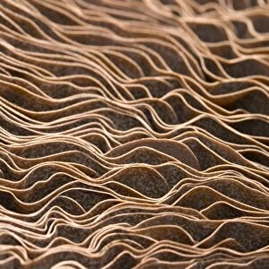
Moss spore capsule, light micrograph

![]()
Wall Art and Photo Gifts from Science Photo Library
Moss spore capsule, light micrograph
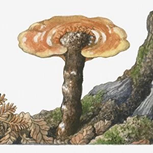
Moss spore capsule, polarised light micrograph. Longitudinal section through a spore capsule from a fire moss (Funaria hygrometrica). The capsules cover is dark green at its base (apophysis), where photosynthetic tissue makes food for the developing spores. The inner capsule (upper centre) is where the spores form. The inner capsule has a lid (operculum, pink), with edges (annulus, bright green) that break down to allow the lid to fall off, releasing the spores. Magnification: x45 when printed at 10 centimetres high
Science Photo Library features Science and Medical images including photos and illustrations
Media ID 6308029
© DR KEITH WHEELER/SCIENCE PHOTO LIBRARY
Bryophyte Bryophytes Bryophytic Cellular Internal Structure Longitudinal Moss Mosses Operculum Orange Orange Background Part Parts Plant Anatomy Polarised Polarized Re Production Reproductive Reproductive Part Reproductive Parts Sporangium Spore Spore Capsule Spores Structural Tissue Annulus Cells Light Micrograph Light Microscope Section Sectioned
EDITORS COMMENTS
This print showcases the intricate beauty of a moss spore capsule, captured under polarized light microscopy. The image provides a longitudinal section view of a spore capsule from the fire moss species known as Funaria hygrometrica. At its base, the dark green cover called apophysis houses photosynthetic tissue responsible for nourishing the developing spores. In the upper center, we can observe the inner capsule where these spores take shape. The pink lid, scientifically referred to as operculum, rests atop the inner capsule and is surrounded by bright green edges called annulus. As part of their reproductive process, these annulus edges gradually break down to allow the lid to detach and release an abundance of tiny spores into their surroundings. Printed at 10 centimeters high with a magnification factor of x45, this image offers us an up-close look at nature's microscopic wonders. It unveils not only the internal structure but also highlights various botanical elements such as cells and tissues that play crucial roles in moss reproduction. With its vibrant orange background contrasting against delicate shades of green and pink, this photograph serves as a testament to both scientific exploration and artistic appreciation for plant anatomy.
MADE IN THE USA
Safe Shipping with 30 Day Money Back Guarantee
FREE PERSONALISATION*
We are proud to offer a range of customisation features including Personalised Captions, Color Filters and Picture Zoom Tools
SECURE PAYMENTS
We happily accept a wide range of payment options so you can pay for the things you need in the way that is most convenient for you
* Options may vary by product and licensing agreement. Zoomed Pictures can be adjusted in the Cart.