Home > Arts > Artists > P > those present
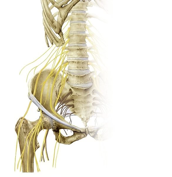
Right hip and nerve plexus, artwork C016 / 6809
![]()

Wall Art and Photo Gifts from Science Photo Library
Right hip and nerve plexus, artwork C016 / 6809
Right hip and nerve plexus. Artwork of the nerves (yellow) and bones of the right hip, seen from the front. This is the lumbar plexus, itself forming part of the lumbosacral plexus, including lumbar and sacral nerves. The main hip nerves present here are those shown passing over or close to the head of the femur. From right to left, these are: the sciatic nerve, the femoral nerve, and the lateral femoral cutaneous nerve. For the left hip, see C016/6808
Science Photo Library features Science and Medical images including photos and illustrations
Media ID 9244873
© D & L GRAPHICS / SCIENCE PHOTO LIBRARY
Anterior Backbone Bones Bundle Femoral Femoral Nerve Femur Frontal Iliac Joint Lower Back Lumbar Plexus Lumbosacral Plexus Nerve Nerve Plexus Nerves Neural Pelvic Sacral Sacrum Sciatic Nerve Skeletal Vertebral Column Neurological Neurology Pelvis
FEATURES IN THESE COLLECTIONS
> Arts
> Artists
> P
> those present
EDITORS COMMENTS
This print showcases the intricate details of the right hip and nerve plexus, providing a fascinating glimpse into the complex anatomy of our bodies. Against a clean white background, this illustration beautifully highlights both the nerves (depicted in vibrant yellow) and bones that make up this vital region. The lumbar plexus takes center stage here, forming part of the larger lumbosacral plexus which includes both lumbar and sacral nerves. The main focus is on the prominent hip nerves that traverse over or near the head of the femur. From right to left, these crucial pathways include: the sciatic nerve, responsible for transmitting signals from lower limbs; followed by the femoral nerve, essential for movement and sensation in thigh muscles; finally culminating with the lateral femoral cutaneous nerve which provides sensory information to parts of your outer thigh. This artwork not only serves as an educational tool but also emphasizes how interconnected our skeletal system is with our neurological network. With its detailed depiction of various structures such as pelvis, vertebral column, sacrum, and even lower back components like iliac bones - it offers a comprehensive view into this intricate web. Created by D & L GRAPHICS for Science Photo Library's collection, this print captures both scientific accuracy and artistic finesse. It invites viewers to appreciate not just their own body's complexity but also ignites curiosity about human biology at large.
MADE IN THE USA
Safe Shipping with 30 Day Money Back Guarantee
FREE PERSONALISATION*
We are proud to offer a range of customisation features including Personalised Captions, Color Filters and Picture Zoom Tools
SECURE PAYMENTS
We happily accept a wide range of payment options so you can pay for the things you need in the way that is most convenient for you
* Options may vary by product and licensing agreement. Zoomed Pictures can be adjusted in the Cart.

