Home > Europe > United Kingdom > Scotland > Moray > Keith
Compact bone, light micrograph

![]()
Wall Art and Photo Gifts from Science Photo Library
Compact bone, light micrograph
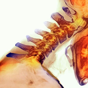
Compact bone. Polarised light micrograph of a transverse section through compact bone tissue, showing Haversian canals (circular regions). The concentric rings surrounding the Haversian canals are called lamellae. The canals run along the long skeletal bones, and contain blood vessels, lymphatic vessels, and nerves. The lamellae are made of compacted collagen fibres and minerals produced by bone-forming cells that are called osteoblasts. The osteoblasts become trapped in the bone matrix at sites called lacunae (small dark spots spaced throughout). Magnification: x103 when printed at 10 centimetres across
Science Photo Library features Science and Medical images including photos and illustrations
Media ID 6278945
© DR KEITH WHEELER/SCIENCE PHOTO LIBRARY
Bony Calcified Calcium Canaliculi Canaliculus Collagen Fibre Collagen Fibres Compact Bone Cross Section Haversian Canal Histological Histology Lacuna Lacunae Lamella Lamellae Matrix Osteocyte Osteocytes Osteology Polarised Polarized Tissue Transverse Light Micrograph Light Microscope Section Sectioned
FEATURES IN THESE COLLECTIONS
> Arts
> Artists
> L
> polarized light
> Europe
> United Kingdom
> Scotland
> Moray
> Keith
> Science Photo Library
> Specialist Imaging
EDITORS COMMENTS
This print showcases the intricate structure of compact bone tissue under a polarised light microscope. The transverse section reveals Haversian canals, circular regions that serve as vital conduits within long skeletal bones. These canals house an extensive network of blood vessels, lymphatic vessels, and nerves, ensuring proper nourishment and communication throughout the bone. The concentric rings surrounding the Haversian canals are known as lamellae. Composed of tightly packed collagen fibers and minerals produced by osteoblasts (bone-forming cells), these lamellae provide strength and support to the bone matrix. Within this matrix, small dark spots called lacunae can be observed where osteoblasts become trapped. With a magnification of x103 when printed at 10 centimeters across, this image offers a detailed glimpse into the fascinating world of human anatomy. It highlights the importance of calcium in maintaining healthy bones while shedding light on various biological processes such as osteology and histology. Expertly captured by Science Photo Library, this photograph is not only visually stunning but also serves as a valuable educational resource for those interested in understanding the complexity and beauty hidden within our bodies' structural framework.
MADE IN THE USA
Safe Shipping with 30 Day Money Back Guarantee
FREE PERSONALISATION*
We are proud to offer a range of customisation features including Personalised Captions, Color Filters and Picture Zoom Tools
SECURE PAYMENTS
We happily accept a wide range of payment options so you can pay for the things you need in the way that is most convenient for you
* Options may vary by product and licensing agreement. Zoomed Pictures can be adjusted in the Cart.