Home > Animals > Fishes > S > Sucker
Sheep tick, SEM

![]()
Wall Art and Photo Gifts from Science Photo Library
Sheep tick, SEM
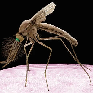
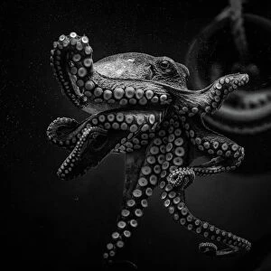
Sheep tick. Coloured scanning electron micrograph (SEM) of a sheep tick (Ixodes ricinus). The ticks mouthparts are between its two front legs. This arachnid is the principal vector of Lyme disease, which is caused by the bacterium Borrelia burgdorferi, in Europe. It is common in the damp underbrush of European woods and attacks various domestic and wild animals, including dogs and humans. Lyme disease causes skin lesions, neurological and cardiac abnormalities, and recurrent arthritis
Science Photo Library features Science and Medical images including photos and illustrations
Media ID 6465949
© STEVE GSCHMEISSNER/SCIENCE PHOTO LIBRARY
Arachnid Arachnida Blood Feeder Blood Sucker Disease Vector Ecto Parasite Ixodes Ricinus Legs Lyme Disease Mouth Parasite Parasitic Sheep Tick False Coloured
FEATURES IN THESE COLLECTIONS
> Animals
> Fishes
> S
> Sucker
> Animals
> Mammals
> Bovidae
> Sheep
> Arts
> Still life artwork
> Still life art
> Nature-inspired artwork
> Arts
> Still life artwork
> Nature-inspired art
> Arts
> Portraits
> Still life artwork
> Nature-inspired artwork
> Arts
> Realistic drawings
> Still life artwork
> Fine art
> Europe
> Related Images
> Popular Themes
> Sheep
> Science Photo Library
> Specialist Imaging
EDITORS COMMENTS
This print showcases the intricate details of a sheep tick, captured using a scanning electron microscope. The vibrant colors bring to life this arachnid, known as Ixodes ricinus, which plays a significant role in transmitting Lyme disease throughout Europe. Positioned between its two front legs are the ticks' mouthparts, highlighting its parasitic nature. Found commonly in the damp underbrush of European woods, this blood-sucking creature targets both domestic and wild animals including dogs and humans. Its ability to transmit Borrelia burgdorferi bacteria makes it the principal vector for Lyme disease. This debilitating illness can lead to skin lesions, neurological and cardiac abnormalities, as well as recurrent arthritis. The image not only provides an up-close look at this fascinating zoological specimen but also serves as a reminder of the delicate balance within our natural world. By studying these parasites through advanced microscopy techniques like SEM, scientists gain valuable insights into their biology and behavior. Displayed against a clean background with cut-out precision, this false-colored photograph allows us to appreciate the beauty hidden within even the tiniest creatures that coexist with us on Earth. It is through such scientific exploration that we deepen our understanding of nature's intricacies while striving towards better prevention and treatment strategies for diseases transmitted by these ecto-parasites.
MADE IN THE USA
Safe Shipping with 30 Day Money Back Guarantee
FREE PERSONALISATION*
We are proud to offer a range of customisation features including Personalised Captions, Color Filters and Picture Zoom Tools
SECURE PAYMENTS
We happily accept a wide range of payment options so you can pay for the things you need in the way that is most convenient for you
* Options may vary by product and licensing agreement. Zoomed Pictures can be adjusted in the Cart.