Home > Science > SEM
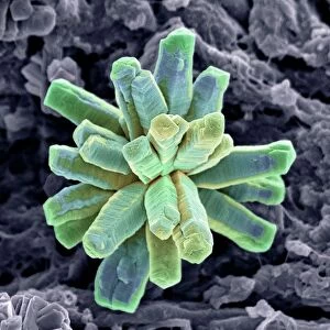
Ovarian follicle, SEM

![]()
Wall Art and Photo Gifts from Science Photo Library
Ovarian follicle, SEM
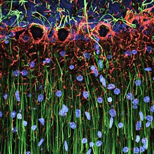
Ovarian follicle. Coloured scanning electron micrograph (SEM) of a fracture through a secondary follicle in the ovary. The oocyte (developing egg) is orange and its central nucleus is darker orange. Surrounding the oocyte is the zona pellucida (green) and a layer of follicular cells known as the corona radiata (purple). Magnification: x1350 when printed at 10 centimetres wide
Science Photo Library features Science and Medical images including photos and illustrations
Media ID 6322439
© STEVE GSCHMEISSNER/SCIENCE PHOTO LIBRARY
Corona Radiata Developing Female Reproductive System Maturing Nucleus Oocyte Ovarian Ovary Ovum Re Production Reproductive Secondary Follicle Zona Pellucida False Coloured
FEATURES IN THESE COLLECTIONS
> Science Photo Library
> Specialist Imaging
EDITORS COMMENTS
This print showcases the intricate beauty of an ovarian follicle, captured through a scanning electron microscope (SEM). The image reveals a fracture through a secondary follicle in the ovary, offering us a glimpse into the fascinating world of female reproductive anatomy. At the center of attention is the developing egg, known as the oocyte. Its vibrant orange hue draws our eyes towards it, while its central nucleus appears darker and more pronounced. Surrounding this precious oocyte is a protective layer called the zona pellucida, depicted here in striking green. This translucent barrier shields and nurtures the developing egg on its journey towards maturity. Adding to this mesmerizing composition are delicate purple hues representing another layer of cells known as corona radiata. These follicular cells form an essential part of the ovarian structure and play vital roles in supporting and nourishing the growing oocyte. With a magnification level set at x1350 when printed at 10 centimeters wide, every minute detail becomes apparent. This false-colored SEM image not only offers aesthetic appeal but also serves as an invaluable tool for studying reproduction biology and understanding various stages within this complex process. Science Photo Library has once again provided us with an extraordinary visual representation that merges artistry with scientific exploration.
MADE IN THE USA
Safe Shipping with 30 Day Money Back Guarantee
FREE PERSONALISATION*
We are proud to offer a range of customisation features including Personalised Captions, Color Filters and Picture Zoom Tools
SECURE PAYMENTS
We happily accept a wide range of payment options so you can pay for the things you need in the way that is most convenient for you
* Options may vary by product and licensing agreement. Zoomed Pictures can be adjusted in the Cart.