Home > Science > SEM
Synapse, SEM C018 / 0122
![]()

Wall Art and Photo Gifts from Science Photo Library
Synapse, SEM C018 / 0122
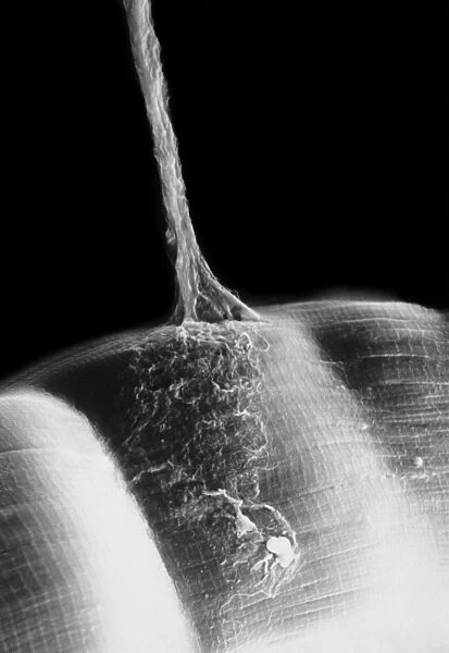
Synapse. Scanning electron micrograph (SEM) of a neuromuscular junction showing a motor neurone (vertical line) terminating on skeletal muscle fibres (across bottom frame). The axon of a motor neurone terminates in several branching fibres, each of which ends in an end plate, a cluster of small swellings, or boutons. When activated, the boutons release neurotransmitter chemicals from small vesicles. The neurotransmitters diffuse across the gap, or synaptic cleft, separating the axon and muscle and bind with receptors in the muscle cell. Magnification: x940 when printed at 10 centimetres tall
Science Photo Library features Science and Medical images including photos and illustrations
Media ID 9237031
© CNRI/SCIENCE PHOTO LIBRARY
Axon Fibre Magnified Image Microscopic Photos Motor Motor Neurone Nerve Nerve Cell Neurone Subjects Synapse Transmitting Bouton Cells Muscle Cell Nervous System Neurological Neurology
FEATURES IN THESE COLLECTIONS
EDITORS COMMENTS
This print titled "Synapse, SEM C018 / 0122" offers a mesmerizing glimpse into the intricate world of our nervous system. Captured through a scanning electron microscope (SEM), this image showcases a neuromuscular junction where a motor neurone connects with skeletal muscle fibres. The vertical line represents the motor neurone's axon, which branches out into several fibres that terminate in small swellings known as boutons. These boutons play a crucial role in transmitting signals between nerve cells and muscles. When activated, they release neurotransmitter chemicals from tiny vesicles. The synaptic cleft, depicted as the gap separating the axon and muscle cell, is where these neurotransmitters diffuse across to bind with receptors in the muscle cell. This binding triggers various physiological responses within the muscle fiber. At x940 magnification when printed at 10 centimeters tall, this monochrome image reveals astonishing details that are otherwise invisible to the naked eye. It serves as a testament to both the complexity and beauty found within our own bodies. With its focus on motor neurons, muscles, synapses, and other elements of neurology and biology, this photograph provides valuable insights for researchers studying subjects related to human anatomy or neurological disorders. Its scientific significance is further enhanced by being part of CNRI/SCIENCE PHOTO LIBRARY's collection of microscopic photos captured using advanced imaging techniques like SEMs.
MADE IN THE USA
Safe Shipping with 30 Day Money Back Guarantee
FREE PERSONALISATION*
We are proud to offer a range of customisation features including Personalised Captions, Color Filters and Picture Zoom Tools
SECURE PAYMENTS
We happily accept a wide range of payment options so you can pay for the things you need in the way that is most convenient for you
* Options may vary by product and licensing agreement. Zoomed Pictures can be adjusted in the Cart.

