Home > Animals > Mammals > Muridae > Fortior
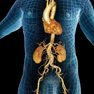
Ischaemia, digital angiogram

![]()
Wall Art and Photo Gifts from Science Photo Library
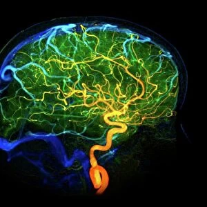
Ischaemia, digital angiogram
image d angiographie numerisee( non soustraite) de la main gauche, en vue de face, par catheterisme hyperselectif des arteres du membre superieur gauche, sous anasthesie generale, chez un patient age de 40 ans, presentant une ischemie de la partie distale du 2A doigt :presence d un aspect irregulier de la partie proximale de l artere ulnaire, avec thrombose focale et thrombose longitudinale non stenosantes, presence d une occlusion de l artere interosseuse entre le 2A'&3A rayon, les arteres colaterales distales du 2A doigt sont occluses et presentent des petites images endoluminales evocatrices de thrombus ( tableau d arterite )
Science Photo Library features Science and Medical images including photos and illustrations
Media ID 10936367
© ZEPHYR/SCIENCE PHOTO LIBRARY
Angiogram Angiography Arterial Arteries Blockage Blocked Blocking Blood Flow Bones Clot Clots Clotted Clotting Colored Diagnosis Diagnostic Imaging Fingers Forties Hand Injured Injury Ischaemia Ischaemic Ischemia Metacarpal Metacarpals Obstructed Obstruction Occlusion Proximal Radiography Radiological Radiology Restricted Restriction Thrombosis Vascular Vessels X Ray Machine Xray Abnormal Artery Blood Supply Blood Vessel Condition Disorder Index Finger Unhealthy
FEATURES IN THESE COLLECTIONS
> Animals
> Mammals
> Muridae
> Fortior
> Arts
> Minimalist artwork
> Monochrome artwork
> Fine art
> Arts
> Minimalist artwork
> Monochrome artwork
> Monochrome paintings
> Arts
> Street art graffiti
> Digital art
> Digital paintings
EDITORS COMMENTS
This print showcases a digital angiogram of the left hand, taken through hyperselective catheterization of the arteries in the upper left limb. The patient, a 40-year-old individual suffering from ischemia in the distal part of their second finger (2A), underwent this procedure under general anesthesia. Upon examination, irregularities were observed in the proximal section of the ulnar artery, accompanied by focal and non-stenosing longitudinal thromboses. Further analysis revealed an occlusion in the interosseous artery between rays 2A and 3A, with occluded distal collateral arteries displaying small endoluminal images suggestive of thrombus formation. This complex presentation suggests a potential diagnosis of arteritis. The monochrome image effectively highlights these abnormalities within the vascular system while providing valuable insights into blood flow restriction and vessel blockages. Radiological imaging techniques like digital angiography play a crucial role in diagnosing such conditions accurately. This photograph serves as a powerful reminder of both our intricate vascular network and its vulnerability to injury or disease. It underscores how medical advancements continue to enable healthcare professionals to identify and understand various disorders affecting blood supply within our bodies.
MADE IN THE USA
Safe Shipping with 30 Day Money Back Guarantee
FREE PERSONALISATION*
We are proud to offer a range of customisation features including Personalised Captions, Color Filters and Picture Zoom Tools
SECURE PAYMENTS
We happily accept a wide range of payment options so you can pay for the things you need in the way that is most convenient for you
* Options may vary by product and licensing agreement. Zoomed Pictures can be adjusted in the Cart.