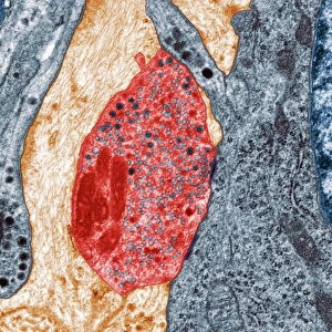
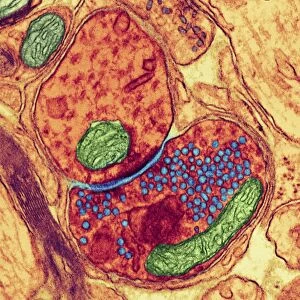
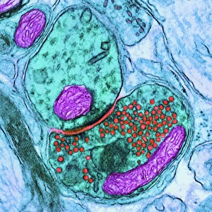
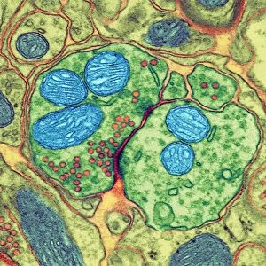
Coloured TEM of a nerve synapse

![]()
Wall Art and Photo Gifts from Science Photo Library
Coloured TEM of a nerve synapse
Synapse. Coloured Transmission Electron Micrograph (TEM) of a synapse. These transmit one-way signals from one nerve cell to the next. The end of the nerve cell (known as a terminal bouton) that transmits the signal is seen at upper centre (green). This is a roughly triangular shape and contains cell-organelles. At centre, a multitude of small circular synaptic vesicles are seen (red) which contain a neurotransmitter. When a signal is transmitted, these vesicles are released and cross the microscopic gap (known as the synaptic cleft) to the next cell (seen at lower right). In this way the nerve signal is passed on from cell to cell. Magnification: x14, 400 at 6x7cm size
Science Photo Library features Science and Medical images including photos and illustrations
Media ID 6448891
© PROF S. CINTI/SCIENCE PHOTO LIBRARY
Nerve Cell Nervous Neurone Neurotransmitter Synapse Synaptic Synaptic Cleft System Vesicles Bouton Cells
MADE IN THE USA
Safe Shipping with 30 Day Money Back Guarantee
FREE PERSONALISATION*
We are proud to offer a range of customisation features including Personalised Captions, Color Filters and Picture Zoom Tools
SECURE PAYMENTS
We happily accept a wide range of payment options so you can pay for the things you need in the way that is most convenient for you
* Options may vary by product and licensing agreement. Zoomed Pictures can be adjusted in the Cart.