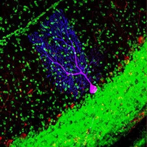
Induced nerve stem cells, micrograph

![]()
Wall Art and Photo Gifts from Science Photo Library
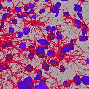
Induced nerve stem cells, micrograph
Induced nerve stem cells. Fluorescence light micrograph of neural (nerve) stem cells that have been created (induced) from human adult skin fibroblast cells by gene manipulation. Tuj1 protein is green, and nestin is blue. Stem cells are pluripotent - they are able to differentiate into any of the 200 cell types in the human body. The type of cell they mature into depends upon the biochemical signals received by the immature cells. This ability makes them a potential source of cells to repair damaged tissue in diseases such as Parkinsons and insulin-dependent diabetes
Science Photo Library features Science and Medical images including photos and illustrations
Media ID 9222927
© SILVIA RICCARDI/SCIENCE PHOTO LIBRARY
Cell Biology Culture Cultured Cytology Derived Fibroblast Fibroblasts Fluorescence Light Micrograph Fluorescent Genes Genetic Genetic Engineering Genome Histological Histology Induced Modification Modified Nerve Cell Neural Neuron Neurone Neurones Neurons Neuroscience Pluripotent Precursor Cell Proteins Skin Cell Stem Cell Therapeutic Treatment Tuj1 Biochemical Biochemistry Cells Genetics Light Microscope Neurological Neurology Protein
EDITORS COMMENTS
This print showcases induced nerve stem cells, offering a glimpse into the incredible potential of gene manipulation in medical science. The micrograph reveals neural stem cells that have been derived from human adult skin fibroblast cells through genetic engineering. In this image, the Tuj1 protein is depicted in vibrant green, while nestin appears as a striking blue hue. These induced nerve stem cells possess pluripotency, meaning they hold the remarkable ability to differentiate into any of the 200 cell types found within the human body. Their fate and specialization are determined by biochemical signals received during their maturation process. This unique characteristic positions them as an invaluable resource for repairing damaged tissues caused by diseases like Parkinson's and insulin-dependent diabetes. The implications of this breakthrough extend far beyond what meets the eye in this fluorescent micrograph. With their potential therapeutic applications, these cultured stem cells offer hope for patients seeking innovative treatments in neurology and other branches of medicine. By harnessing genetic modifications and understanding cellular biology at its core, scientists continue to unlock new frontiers in regenerative medicine. Science Photo Library presents this awe-inspiring image not only as a testament to scientific progress but also as a reminder of how our understanding of genetics and biochemistry can revolutionize healthcare outcomes for millions worldwide.
MADE IN THE USA
Safe Shipping with 30 Day Money Back Guarantee
FREE PERSONALISATION*
We are proud to offer a range of customisation features including Personalised Captions, Color Filters and Picture Zoom Tools
SECURE PAYMENTS
We happily accept a wide range of payment options so you can pay for the things you need in the way that is most convenient for you
* Options may vary by product and licensing agreement. Zoomed Pictures can be adjusted in the Cart.