Home > Animals > Mammals > Rodents
Hippocampus brain tissue

![]()
Wall Art and Photo Gifts from Science Photo Library
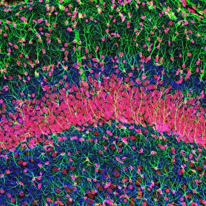
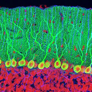
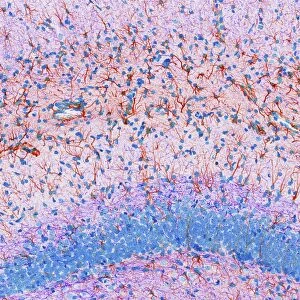
Hippocampus brain tissue
Hippocampus brain tissue. Light micrograph of a sagittal (side view) section through the hippocampus of a rats brain showing the nerve cells within it. The hippocampus is a structure of the limbic system of the brain. There is a hippocampus in each cerebral hemisphere, and they are thought to be responsible for spatial awareness and the formation of memories. Different structures have been stained different colours: the nuclei of cells are orange and neurofilaments are blue. Neurofilaments are proteins found in the axons (output processes) of nerve cells
Science Photo Library features Science and Medical images including photos and illustrations
Media ID 6335764
© THOMAS DEERINCK, NCMIR/SCIENCE PHOTO LIBRARY
Animal Tissue Hippocampus Histological Histology Nerve Cell Neurofilament Neurofilaments Neuron Neurone Neurones Neurons Neuroscience Nuclei Nucleus Rodent Sagittal Slice Stained Tissue Brain Cells Light Micrograph Light Microscope Nervous System Neurological Neurology Section Sectioned
MADE IN THE USA
Safe Shipping with 30 Day Money Back Guarantee
FREE PERSONALISATION*
We are proud to offer a range of customisation features including Personalised Captions, Color Filters and Picture Zoom Tools
SECURE PAYMENTS
We happily accept a wide range of payment options so you can pay for the things you need in the way that is most convenient for you
* Options may vary by product and licensing agreement. Zoomed Pictures can be adjusted in the Cart.