Home > Popular Themes > Human Body
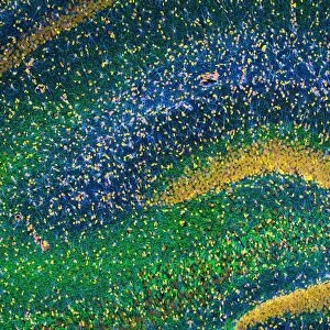
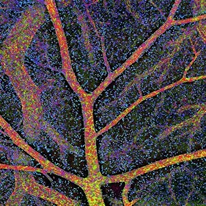
Hippocampus brain tissue

![]()
Wall Art and Photo Gifts from Science Photo Library
Hippocampus brain tissue
Hippocampus tissue. Light micrograph of a sagittal (side view) section through the hippocampus of the brain showing the nerve cells within it. The hippocampus is a structure of the limbic system of the brain. There is a hippocampus in each cerebral hemisphere, and they are thought to be responsible for spatial awareness and the formation of memories. Different structures have been stained different colours: the nuclei of cells are red, neurofilaments are blue and glial cells are green. Neurofilaments are proteins found in the axons (output processes) of nerve cells. Glial cells (neuroglia) are cells that support and nourish nerve cells. Magnification: x160 when printed 10cm wide
Science Photo Library features Science and Medical images including photos and illustrations
Media ID 6448743
© THOMAS DEERINCK, NCMIR/SCIENCE PHOTO LIBRARY
Glia Glial Hippocampus Histological Histology Nerve Cell Neurofilament Neurofilaments Neuroglia Neuron Neurons Nuclei Nucleus Sagittal Slice Brain Cells Light Micrograph Light Microscope Nervous System Neurological Neurology Section Sectioned
EDITORS COMMENTS
This print showcases the intricate beauty of hippocampus brain tissue. In this light micrograph, a sagittal section provides a side view into the hippocampus, which is an essential component of the limbic system in our brains. The nerve cells within this structure are prominently displayed, offering a glimpse into their complex network. The hippocampus plays a crucial role in spatial awareness and memory formation. Each cerebral hemisphere houses its own hippocampus, working together to support these cognitive functions. To distinguish various structures within the tissue, different colors have been used: red for cell nuclei, blue for neurofilaments (proteins found in nerve cell axons), and green for glial cells (neuroglia) that provide nourishment and support to neurons. With a magnification of x160 when printed 10cm wide, this image allows us to appreciate the microscopic details that make up our brain's anatomy. It serves as a reminder of how intricately designed our bodies are at every level. This photograph from Science Photo Library offers valuable insights into biology, anatomy, neurology, and histology. Its depiction of healthy brain tissue provides scientists and medical professionals with invaluable knowledge about the nervous system and its functioning.
MADE IN THE USA
Safe Shipping with 30 Day Money Back Guarantee
FREE PERSONALISATION*
We are proud to offer a range of customisation features including Personalised Captions, Color Filters and Picture Zoom Tools
SECURE PAYMENTS
We happily accept a wide range of payment options so you can pay for the things you need in the way that is most convenient for you
* Options may vary by product and licensing agreement. Zoomed Pictures can be adjusted in the Cart.